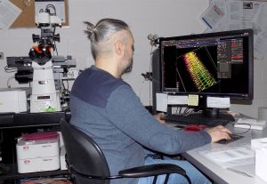
The Nikon A1 Laser Scanning Confocal Microscope is configured on a Nikon Ti automated inverted microscope platform with an XY automated stage and provides up to 6 lines of laser excitation. Laser excitation includes the 405nm, 445nm, 488nm, 514nm, 561nm and 647nm diode laser lines. A hybrid confocal scan head provides both a galvano scanner for high-resolution imaging and a high-speed resonance scanner for live cell imaging. Confocal fluorescence and/or reflection are detected through a single, variable pinhole aperture and are recorded using two standard photomultiplier (PMT) detectors and two high-sensitivity Gallium Arsenide Phosphide (GaAsP) PMT detectors. An independent PMT detector permits collection of brightfield, polarized light, and DIC contrast images. A 32-channel spectral detector with spectral unmixing software is available for separation of overlapping fluorescence emission spectra and/or background autofluorescence. The system is configured with a fully automated XY stage that may be used for automated acquisition of high-resolution large area scans.
The Nikon A1 inverted microscope system is also configured with optics and software for Total Internal Reflection Fluorescence (TIRF) microscopy and 2D/3D super resolution STORM imaging. For these specialized applications, the system is configured with a high-sensitivity Andor iXon DU897 512×512 (16 micrometer pixel) EMCCD camera and a 100x Apo TIRF objective (NA 1.49). A 405/488/561/647 quadruple pass filter cube and a 445/514/561/ triple pass filter cube are available to accommodate a wide range of fluorochromes for these applications. Nikon A1 CLSM
Advanced imaging applications include:
- High-resolution, high-sensitivity confocal imaging in both fluorescence and reflected light modes
- Multi-channel confocal fluorescence imaging, including blue fluorescence (excitation 405 nm) through far red fluorescence (excitation 647 nm)
- 3D rendering and animation
- Co-localization analysis
- Cell counting and image analysis software
- Live cell imaging and quantitative analyses over time
- FRAP and FRET imaging and analysis
- Fluorescence protein imaging, including CFP, GFP, YFP, RFP, and BiFC analysis
- Spectral imaging using a 32-channel spectral detector with 2.5nm, 5nm, and 10nm bandwidth selection
- Transmitted light imaging (brightfield, polarized light, DIC)
- OKO Lab heating/cooling/carbondioxide environmental stage incubator
