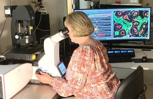
The Leica Stellaris 5 Laser Scanning Confocal Microscope is configured on an automated Leica DMi8 inverted microscope platform with an XY automated stage. Laser excitation is provided by a 405nm diode laser, a 448nm diode laser, and a pulsed supercontinuum white light laser that provides a tunable range of laser excitation from 488nm to 685nm. Fluorescence emissions are spectrally separated through a prism design and isolated through variable slits that provide bandwidths that are freely adjustable in 1nm increments. Four high-sensitivity Power HyD S detectors are available for simultaneous or sequential recording of the confocal fluorescence, with an independent detector provided for the collection of brightfield, polarized light, and DIC images.
The Stellaris 5 offers a range of advanced imaging applications including live cell imaging, 3-dimensional imaging and reconstruction, spectral unmixing for separation of overlapping fluorescence emissions, fluorescence lifetime imaging, targeted photoactivation and photobleaching, imaging stitching for large area scanning, and super resolution imaging.
The Leica Stellaris 5 advanced imaging features and applications include:
- High-resolution, high-sensitivity confocal imaging in both fluorescence and reflected light imaging modes.
- Multi-channel confocal fluorescence imaging through a 410 nm – 850 nm emission detection range.
- Continuous wave 405nm diode and 448nm diode lasers for confocal imaging, as well as targeted photoactivation and photobleaching applications.
- Pulsed supercontinuum white light laser, providing a tunable range of laser excitation from 488nm-685nm in 1 nm increments for confocal imaging.
- TauSense lifetime imaging software (including TauContrast, TauSeparation, TauGating, and TauInteraction) which, in combination with the pulsed white light laser, provides fluorochrome lifetime information that can be used to monitor changes in cellular microenvironments, improve spectral separation of overlapping dyes, and remove unwanted autofluorescence.
- Filter-free, spectral separation of fluorescence emission provided by a prism design.
- Freely adjustable bandwidth detectors that can be adjusted in 1nm increments.
- Four high-sensitivity Power HyD S detectors, providing increased photon detection efficiency in the blue-green range, expanded detection sensitivity up to 850nm, and the option of analog or photon counting modes.
- High quality Plan Apo objectives, including a 10x Plan Apo dry (NA 0.40), 20x Plan Apo dry (NA 0.75), 40x Plan Apo oil (NA 1.30), 63x Plan Apo oil (NA 1.40), 100x Plan Apo oil (NA 1.40), 20x Plan Apo multi-immersion (oil, glycerol, water) (NA 0.75), and 40x Plan Apo water (NA 1.10).
- 3D rendering and animation software.
- Live cell imaging and quantitative analyses over time, including FRAP and FRET imaging and analysis.
- Lambda scanning, with a variable bandwidth selection down to 5 nm, and spectral unmixing for separation of overlapping fluorescence emission spectra and background autofluorescence.
- Navigator mosaic tile scanning for large area scanning and stitching.
- Lightning Super Resolution imaging, providing super resolution up to 120 nm lateral (XY) and 200 nm axial (Z) resolution.
- Transmitted light imaging (brightfield, polarized light, DIC)