The Nikon C2 Laser Scanning Confocal Microscope is configured on Nikon Ni manual upright microscope platform with up to 4 lines of laser excitation, including the 405nm, 488nm, 561nm, and 640nm laser lines. High-resolution confocal fluorescence and/or reflection are detected through a single, variable pinhole aperture and are recorded using three high-sensitivity photomultiplier (PMT) detectors with band pass and long pass emission filters for wavelength selection. An independent PMT detector permits collection of brightfield, phase contrast, polarized light, and DIC contrast images.
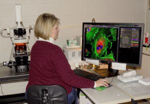 The Nikon C2 CLSM
Advanced imaging applications include:
- High-resolution confocal imaging in both fluorescence and reflected light modes
- Multi-channel confocal fluorescence imaging, including blue fluorescence (excitation 405 nm) through far red fluorescence (excitation 640nm)
- 3D rendering and animation
- Co-localization analysis
- Cell counting and analysis software
- Live cell imaging and quantitative analysis over time
- FRAP and FRET imaging and analysis
- Fluorescence protein imaging, including CFP, GFP, YFP, RFP and BiFC analysis
- Extended Depth of Focus (EDF) analysis, Topography analysis, Z height profiling
- Transmitted light imaging (brightfield, phase contrast, polarized light, DIC)
- DS-Fi2 digital color camera for conventional fluorescence and transmitted light imaging.